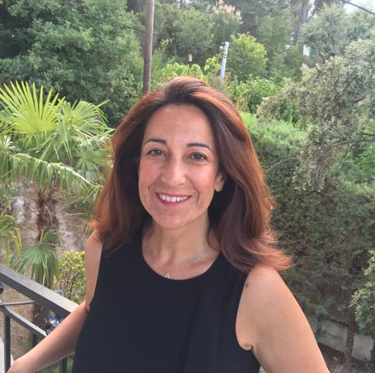
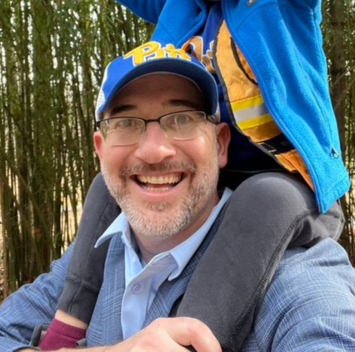
Agenda
Saturday, February 21
| 8:00 AM - 9:00 AM | |
| 9:00 AM - 4:00 PM |
Sunday, February 22
| 8:00 AM - 7:00 PM | |
| 9:00 AM - 12:00 PM | |
| 9:00 AM - 4:00 PM | |
| 1:00 PM - 4:00 PM | |
| 2:00 PM - 4:00 PM | |
| 4:00 PM - 4:30 PM | |
| 4:30 PM - 5:45 PM | |
| 6:00 PM - 7:00 PM | |
| 7:00 PM - 8:30 PM |
Monday, February 23
| 6:00 AM - 7:00 AM | |
| 7:00 AM - 4:30 PM | |
| 7:15 AM - 8:15 AM | |
| 8:30 AM - 9:05 AM | |
| 9:15 AM - 10:35 AM | |
| 10:35 AM - 11:00 AM | |
| 11:00 AM - 12:20 PM | |
| 12:30 PM - 1:30 PM | |
| 1:40 PM - 3:00 PM | |
| 3:15 PM - 4:30 PM | |
| 4:30 PM - 6:30 PM | |
| 6:30 PM - 7:30 PM | |
| 7:30 PM - 8:30 PM |
Tuesday, February 24
| 7:00 AM - 4:30 PM | |
| 7:15 AM - 8:15 AM | |
| 8:30 AM - 9:05 AM | |
| 9:15 AM - 10:35 AM | |
| 10:35 AM - 11:00 AM | |
| 11:00 AM - 12:20 PM | |
| 12:30 PM - 1:30 PM | |
| 1:40 PM - 3:00 PM | |
| 3:15 PM - 4:30 PM | |
| 4:30 PM - 6:30 PM | |
| 6:30 PM - 7:30 PM | |
| 7:00 PM - 8:30 PM |
Wednesday, February 25
| 7:00 AM - 11:00 AM | |
| 7:15 AM - 8:15 AM | |
| 8:30 AM - 9:05 AM | |
| 9:15 AM - 10:35 AM | |
| 10:35 AM - 11:00 AM | |
| 11:00 AM - 1:00 PM |
Saturday, February 21
| 8:00 AM - 9:00 AM - Registration | |||
#
Saturday Short Course Registration |
|||
| 9:00 AM - 4:00 PM - Short Course | |||
#
Introduction to Machine Learning and Artificial Intelligence for Proteomics Data AnalysisMachine learning-and correspondingly, artificial intelligence-has become a dominant technology for data-intensive discovery in nearly all scientific domains. Today, almost all biomedical research employs machine learning techniques to derive new knowledge from complex biological data. This course will introduce the fundamentals of machine learning with a specific focus on the analysis of proteomics data, through a series of lectures and hands-on labs. The primary goal of this course is to promote data literacy for people new to machine learning. Students who complete the course will be equipped to critically evaluate uses of machine learning in scientific literature, recognize common pitfalls, perform basic machine learning analyses, and know when to consult a machine learning expert-and how to communicate with them. The topics that will be covered include: - What is machine learning? - Types of machine learning tasks - The bias-variance trade-off - Machine learning model evaluation - Linear models and logistic regression - Decision tree-based models - Neural networks and artificial intelligence Course participants will: - Recognize when machine learning methods may be beneficial for their research. - Identify common pitfalls in the application of machine learning methods. - Gain confidence to provide constructive feedback for applications of machine learning in the manuscripts they review. - Evaluate the strengths and weaknesses of machine learning approaches presented in the scientific literature. - Gain familiarity with additional resources to deepen their understanding of machine learning. Prerequisites: All participants will need to bring a laptop to perform the lab exercises during the course. A familiarity with proteomics data analysis is recommended. No machine learning experience is needed. No programming experience is necessary; however, some programming experience in any language is recommended and we will introduce Python as part of the course. Presented By:
|
|||
#
Top-Down For the MassesThe goal of this course is for you to gain the ability to produce and interpret publishable (ideally world-class), deep sequence coverage, Top-Down Mass Spectrometry (TDMS) data. All learners, including current TDMS practitioners, will learn recent advances in internal fragmentation assignment that avoid pervasive false-positive and false-negative assignments and often improve sequence coverage by an order of magnitude. This course has the following learning objectives: 1) Recognize problems well-suited for TDMS. 2) Design and perform TDMS experiments best suited for your particular sample, scientific question, and instrumentation. You will improve mastery using problem sets where you design sample preparation, fractionation, and MS methods best suited for particular preparations and scientific problems. This marks the end of morning session. 3) Interpret TDMS data, including internal fragments, manually and using free software (bring a Windows based computer if you can). 4) Evaluate your interpretation skills and improve them as needed. Here you will be given spectra to interpret, enough time to enjoy the experience, and will then compare your work to "gold-standard" analysis validated by established TDMS laboratories. 5) Avoid common pitfalls in TDMS experiments and data analysis. For those new to TDMS or even MS, basic spectral interpretation skills and MS operational principles will be taught briefly. If you're uncertain of being adequately prepared but are dedicated to becoming so, online introductory tutorials in spectral interpretation and instrumentation will provided in advance. Presented By:
|
|||
#
Quantitative Data Analysis for Proteomics - Part 1This course will cover both theory and practice for analyzing quantitative proteomics data. The course will be a mixture of both "lecture" and "lab" portions. The "lectures" will discuss best practices, considerations, and pitfalls for interpreting label-free and labeled measurements, including those produced by DDA, DIA, PRM, TMT/iTRAQ, and SILAC methods. The "labs" will give students a chance to explore and analyze raw data from different instrument platforms with open-source tools using Windows laptops that they either bring with them or have remote access to. While participants are highly recommended to bring their own computers to get the full experience, participants without Windows laptops will be able to join up as teams to analyze datasets. The goal of this course is to give students a foundation for how data analysis tools work and to build an intuition for what data analysis challenges lurk in their datasets. A portion of the class will be devoted to "office hours" where students will get a chance to discuss their specific quantitative proteomics challenges. Audience: proteomics researchers at all levels that want to learn what common proteomics data analysis tools/methods are doing, how they work, and where they break down. Presented By:
|
|||
Sunday, February 22
| 8:00 AM - 7:00 PM - Registration | |||||||
#
Registration and Information Desk |
|||||||
| 9:00 AM - 12:00 PM - Short Course | |||||||
#
Single Cell to Spatial ProteomicsThe purpose of this half day course is to gain fundamental knowledge on state-of-the-art single cell to spatialomics proteomic practices, enable discussion on the reality of single cell to spatialomics practices, and build a scholarly support network for proteomic research. The course will be taught in three case studies spanning single cell proteomics by LC-MS/MS, combining proteomics with other 'omic workflows, and leveraging spatial proteomics for studies on the tissue microenvironment. Lectures by leading scientists will present case studies on single cell to spatialomics with discussion and perspectives to include: - Considerations for sample storage and preparation - Deployment of internal standards - Proteomic analysis strategies: Instrumentation and data analysis - Multiomic capabilities: Integrating with current workflows The workshop is for researchers within the proteomic community who wish to update or inform themselves on current trends in single cell to spatial proteomics. Study Topics and Lecturers: - Discovery of Novel Cell Phenotypes Using Single Cell Proteomics: A Case Study on Aortic Aneurysm. Discussion led by Sarah Jessica Parker, Cedars Sinai. This case study focuses on a step-by-step discussion of single sample preparation to data analysis and integration with spatial proteomics. - Deploying Multiomic Analysis with Proteomics Approaches: Studies on Aging, Osteoarthritis, and Age-Related Diseases. Discussion led by Birgit Schilling, Buck Institute. The lecture focuses on studies integrating multiomic workflows with existing proteomic and spatial proteomic workflows. - Implementing Multiomic Studies for Spatial Pathology from One of a Kind Clinically Archived Breast Cancer Tissues. Discussion led by Peggi Angel, Medical University of South Carolina. The lecture focuses leveraging multiomic workflows targeting glycomics, extracellular, and cellular proteomics on single tissue sections. Presented By:
|
|||||||
| 9:00 AM - 4:00 PM - Short Course | |||||||
#
LC-MS 101: Everything You Ever Wanted to Know But Were Afraid to AskMost proteomics measurements utilize liquid chromatography separations coupled with high resolution/mass accuracy mass spectrometry. With these technologies maturing, it becomes possible to collect data while treating the LC-MS platform as a black box, not fully understanding what's going on inside. This practical lecture-style short course will be divided into three topics: separations, ionization and mass measurement. The separations portion will delve into the basics of LC separations, primarily focusing on reversed phase LC but also briefly touching on other modes. We will discuss sample loading and injection strategies, column dimensions, throughput and flow rate considerations. We will utilize some common free software tools to optimize our separations within the constraints of specific instrument capabilities, and students will be invited to follow along with these software tools on their own as desired. We will then describe electrospray ionization and practical considerations to increase robustness, stability and sensitivity. For the mass spectrometry portion, we will describe the ion path and ion optics encountered in modern mass spectrometers, the (very) basics of mass spectrum interpretation and the mass analyzers commonly encountered in proteomics. The course will be highly interactive such that students will be able to get their own questions answered. The target audience is users of LC-MS-based proteomics technologies who either need a refresher or who have never had formal instruction on the use of the instrumentation. Presented By:
|
|||||||
#
Quantitative Data Analysis for Proteomics - Part 2This course will cover both theory and practice for analyzing quantitative proteomics data. The course will be a mixture of both "lecture" and "lab" portions. The "lectures" will discuss best practices, considerations, and pitfalls for interpreting label-free and labeled measurements, including those produced by DDA, DIA, PRM, TMT/iTRAQ, and SILAC methods. The "labs" will give students a chance to explore and analyze raw data from different instrument platforms with open-source tools using Windows laptops that they either bring with them or have remote access to. While participants are highly recommended to bring their own computers to get the full experience, participants without Windows laptops will be able to join up as teams to analyze datasets. The goal of this course is to give students a foundation for how data analysis tools work and to build an intuition for what data analysis challenges lurk in their datasets. A portion of the class will be devoted to "office hours" where students will get a chance to discuss their specific quantitative proteomics challenges. Audience: proteomics researchers at all levels that want to learn what common proteomics data analysis tools/methods are doing, how they work, and where they break down. Presented By:
|
|||||||
#
Capturing the Stable and Transient Protein-Protein InteractionsThis course will introduce various proteomics techniques for studying stable and transient protein-protein interactions, including affinity purification, proximity labeling, and cross-linking methods. The aim is to provide practical tips on study design, experimental optimization, data analysis, and troubleshooting for protein interaction studies. Protein interaction experiments are often challenged by nonspecific binding, experimental variability, contamination, false discoveries, and data analysis bottlenecks. This course will provide practical strategies to address these challenges and optimize experimental design. Instructors with expertise in affinity purification, proximity labeling, and cross-linking techniques will present real-life examples of problems, common pitfalls, and effective solutions. Through both successful and failed experimental examples, attendees will gain insight into best practices for refining protocols, improving data reliability, and overcoming computational hurdles. An interactive discussion session will also be included, giving attendees the opportunity to share their experience and challenges in protein interaction experiments. Presented By:
|
|||||||
| 1:00 PM - 4:00 PM - ECR Event | |||||||
#
ECR Mentoring DayThe US HUPO 2026 Early Career Researcher (ECR) Mentorship Day is back! Join a dynamic, community-driven experience designed to empower trainees, postdocs, and ECRs to thrive in proteomics and beyond. This event brings together inspiring senior researchers for interactive, candid discussions on career strategy, professional development, and the life of a scientist. Expect practical insights, real-world perspectives, and a welcoming environment primed for meaningful and lasting connections to unfold. Stay tuned for exciting announcements on speakers and session themes. |
|||||||
| 2:00 PM - 4:00 PM - Session | |||||||
#
President's Lalapooloza |
|||||||
| 4:00 PM - 4:30 PM - Break | |||||||
#
Break on Own |
|||||||
| 4:30 PM - 5:45 PM - Evening Workshops | |||||||
#
Evening Workshop: Targeting Diabetes and Obesity: MS-based Assays that Inform Clinical Research Transferability |
|||||||
#
Evening Workshop: Proteomics in (Bio)Pharma Industry Part 1This workshop aims to bridge the gap between proteomics research in academic and industrial settings, with a specific focus on the (bio)pharma industry. The workshop will consist of two parts. On day 1 Jan will highlight the unique features, challenges, and opportunities of conducting proteomics work in an industrial setting, where the focus is on delivering medicines for patients in need - while balancing risk, impact and timelines. The day 1 will start with a brief introduction to the drug discovery process, highlighting the three different stages - discovery, pre-clinical, and clinical phases - and how proteomics can play a role in each of them. The main part of the workshop will consist of facilitated discussions on two key topics. Firstly, the availability of opportunities for students to understand scientific work in industrial settings will be explored. Secondly, strategies for demonstrating the impact of proteomics in the (bio)pharma industry will be discussed. Presented By:
|
|||||||
| 6:00 PM - 7:00 PM - Plenary Session | |||||||
#
Opening Plenary SessionDeveloping microbiome-directed therapeutics for treating childhood undernutritionHuman postnatal development is typically viewed from the perspective of our ‘human’ organs. As we come to appreciate how our microbial communities are assembled following birth, there is an opportunity to determine how this microbial facet of our developmental biology is related to healthy growth as well as to the risk for and manifestations of disorders that produce abnormal growth. We are testing the hypothesis that perturbations in the normal development of the gut microbiome are causally related to childhood undernutrition, a devastating global health problem whose long-term sequelae, including stunting, neurodevelopmental abnormalities, plus metabolic and immune dysfunction, remain largely refractory to current therapeutic interventions. The journey to preclinical proof-of-concept, and the path forward to clinical proof-of-concept emphasize the opportunities as well as the experimental, analytic and other challenges encountered when developing microbiota-directed therapeutics. Presented By:
|
|||||||
| 7:00 PM - 8:30 PM - Welcome Reception | |||||||
#
Welcome Reception with ExhibitorsSponsored By: SCIEX  |
|||||||
Monday, February 23
| 6:00 AM - 7:00 AM - Social Activity | |||||||||||||||||
#
Sunrise Run/WalkStart your US HUPO 2026 experience on the move with the Sunrise Run-a refreshing, informal morning run designed to energize your body and mind before a full day of science. Join fellow attendees as we take in the early light, connect beyond the conference, and set a positive pace for the day ahead. All abilities are welcome! Note: Headlamps are recommended, as the run will start shortly before sunrise. Please run in pairs or groups for safety! Each lap is ~1.6 miles, so two laps is approximately a 5k run. Meet in Lobby 10 minutes before 6:00 am. |
|||||||||||||||||
| 7:00 AM - 4:30 PM - Registration | |||||||||||||||||
#
Registration and Information Desk |
|||||||||||||||||
| 7:15 AM - 8:15 AM - Sponsored Seminars | |||||||||||||||||
#
Sponsor Breakfast: Multiplexed Detection of Membranous Nephropathy Antigens in Clinical PracticeSponsored By: Affinisep  Affinisep develops and manufactures from A to Z high quality and ready-to-use microelution SPE kits to simplify sample preparation in proteomics workflows, and help you achieve highly reliable results. Our wide range of formats and capacities allow us to cover a broad range of needs, from few samples processed manually, to high throughput processing of hundreds of samples in clinical studies. After a quick overview of Affinisep's kits for sample preparation in MS-based proteomics, we will have the pleasure to welcome Dr. Aaron Storey, who will present his work on the implementation of a membranous nephropathy typing assay on a triple quadrupole mass spectrometer in clinical practice. Membranous nephropathy is a leading cause of nephrotic syndrome, driven by an immune response against a target autoantigen. Over 30 target antigens have been described in membranous nephropathy to date, creating the need for multiplex approaches for detection. Dr. Storey and his team developed a multi-reaction monitoring-based assay for the clinical mass spectrometry laboratory for determination of an antigen type in nephropathology practice. Immune complex purification and trypsin digestion is performed on a Kingfisher Flex using protein A/G beads for immunoprecipitation and SP3 cleanup. Peptides are desalted using Affinisep BioSPE® plates and are quantified by MRM on a TSQ Altis using AQUA Pro Grade II peptides as internal standards. This immunoprecipitation-to-mass spectrometry workflow demonstrated reliable quantitative detection of nineteen membranous nephropathy antigens. The mass spectrometry-enabled workflow had a diagnostic accuracy of 97.2% with a sensitivity of 97.2% and 100% specificity. A series of cases of consecutive biopsies with PLA2R -negative membranous nephropathy, 57.1% had a rare antigen detected by this workflow. Multi-reaction monitoring mass spectrometry following protein A/G immunoprecipitation of membranous nephropathy biopsy tissue can therefore be effectively used for multiplex antigen typing in a clinical nephropathology practice.
|
|||||||||||||||||
#
Sponsor Breakfast: Standardizing and Scaling Automated Workflows for Cutting Edge Proteomics
|
|||||||||||||||||
| 8:30 AM - 9:05 AM - Plenary Session | |||||||||||||||||
#
Donald F. Hunt Distinguished Contribution in Proteomics Award Plenary SessionAdventures in Quantitative ProteomicsSponsored By: Thermo  Over the past two decades, the MacCoss laboratory has pursued the development of quantitative mass spectrometry methods that are accurate, reproducible, and accessible to the broader proteomics community. Our early work focused on stable isotope labeling strategies, which enabled comprehensive quantitative comparisons of protein abundance across biological conditions. These studies established principles for isotope-based quantification and demonstrated both the promise and challenges of achieving reproducible quantitative measurements in complex biological samples. Recognizing limitations in discovery proteomics for hypothesis-driven research, we focused on the development and application of targeted mass spectrometry approaches. This work emphasized selected reaction monitoring (SRM) and parallel reaction monitoring (PRM) methods that provide sensitive, specific, and reproducible quantification of predetermined protein targets. To make these methods broadly accessible, we developed Skyline, an open-source platform that supports vendor-neutral method development, data analysis, and result sharing. Skyline has become a cornerstone tool for the quantitative proteomics community, enabling laboratories worldwide to implement targeted proteomics workflows. Our laboratory was an early adoptor of data-independent acquisition (DIA) mass spectrometry. We were proponents of 'peptide-centric' analyses of proteomics data as an alternative to 'spectrum-centric' analysis and recognized the ability of DIA to combine the strengths of untargeted discovery methods with the quantitative reproducibility of targeted approaches. Our efforts have worked to improve the selectivitity and specificity of the methodology, minimize false discoveries, reduce challenges with missing data, and improved the visualization of the underlying extracted ion chromatograms of DIA data. Throughout these technical developments, we have maintained a strong emphasis on reproducibility and inter-laboratory harmonization. We established quality control strategies, developed standardized workflows, and created tools for comparing and harmonizing quantitative results across different laboratories, instruments, and experimental designs. Panorama, our web-based data repository, facilitates data sharing and collaborative quality assessment. These quantitative proteomics methods have been successfully applied to diverse biomedical questions spanning aging, cancer, cardiovascular disease, diabetes, and neurodegeneration, demonstrating their broad utility for advancing our understanding of human health and disease. Presented By:
|
|||||||||||||||||
| 9:15 AM - 10:35 AM - Parallel Sessions | |||||||||||||||||
#
Parallel Session 01: Cancer Proteomics
|
|||||||||||||||||
#
Parallel Session 02: Advances in Imaging / Spatial ProteomicsSponsored By: Bruker 
|
|||||||||||||||||
| 10:35 AM - 11:00 AM - Break | |||||||||||||||||
#
Coffee BreakSponsored By: SEER  |
|||||||||||||||||
| 11:00 AM - 12:20 PM - Parallel Sessions | |||||||||||||||||
#
Parallel Session 03: Enabling Structural Biology with Proteomics
|
|||||||||||||||||
#
Parallel Session 04: Affinity Proteomics: Without Mass SpectrometrySponsored By: MilliporeSigma 
|
|||||||||||||||||
| 12:30 PM - 1:30 PM - Sponsored Seminars | |||||||||||||||||
#
Sponsor Lunch: From Discovery, Quantitative Validation, and Deep Characterization: Streamlining Biological Insights with the Zenot of SystemSponsored By: SCIEX  Mass spectrometry-based proteomics has long been constrained by fragmented workflows-researchers often rely on separate instruments for deep discovery proteomics, high precision targeted quantification, and specialized post translational modification (PTM) analyses. These siloed approaches introduce challenges in method transfer, reproducibility, training, budget allocation, and overall laboratory efficiency. This session will introduce a new unified instrumentation paradigm enabled by the ZenoTOF 8600 system, an advanced Zeno trap-enabled TOF platform designed to consolidate historically separated workflows into a single, high performance system. Through new method development strategies and workflow comparisons, this talk will demonstrate how unifying multiple proteomic modes on one instrument not only reduces complexity but also accelerates biological insight. Researchers seeking to eliminate multi system bottlenecks, simplify their pipeline, and achieve higher reproducibility across diverse analytical demands will gain a clear understanding of how this next generation platform reshapes the future of mass spectrometry driven proteomics.
|
|||||||||||||||||
#
Sponsor Lunch: Explore the Human Proteome with Depth and ScaleSponsored By: Olink Proteomics  Proteomics and multiomics are reshaping our understanding of human biology, offering unprecedented insights into disease mechanisms and biomarker landscapes. This session will highlight how Olink's Proximity Extension Assay (PEA) technology is uniquely positioned to support population-scale proteomic studies, bridging the gap from discovery research to clinical utility. The use of integrative analysis combining Olink Explore HT with mass spectrometry analysis provides comprehensive data to identify unique protein signatures and to provide data-driven insights that propel precision medicine.
|
|||||||||||||||||
| 1:40 PM - 3:00 PM - Parallel Sessions | |||||||||||||||||
#
Parallel Session 05: Proximity and Interaction Methods
|
|||||||||||||||||
#
Parallel Session 06: Cardiovascular and Pulmonary Disease Proteomics
|
|||||||||||||||||
| 3:15 PM - 4:30 PM - Lightning Session | |||||||||||||||||
#
Lightning Talks - Round 01 |
|||||||||||||||||
| 4:30 PM - 6:30 PM - Poster Session | |||||||||||||||||
#
Poster Session 01 and Exhibitor Viewing |
|||||||||||||||||
| 6:30 PM - 7:30 PM - Evening Workshops | |||||||||||||||||
#
Evening Workshop: Proteomics Show Live from ...Missouri...Join Ben Orsburn and honorary Ben Neely, Dr. Renã Robinson, for a live recording of the single most popular proteomics podcast, THE Proteomics Show. A volunteer attendee of the conference will randomly be selected and interviewed live on stage with audience participation. We'll talk about research, we'll get feedback on the conference, and maybe we'll find out what route led one conference attendee to involvement in the study of the human proteome. Presented By:
|
|||||||||||||||||
#
Evening Workshop: Proteomics in (Bio)Pharma Industry Part 2This workshop aims to bridge the gap between proteomics research in academic and industrial settings, with a specific focus on the (bio)pharma industry. The workshop will consist of two parts. On Day 2 a panel discussion organized by US HUPO Ed & Out Committee will provide broader context with panelists from diverse industrial settings from start-up scene to major pharmaceutical companies. Full list of panelists will be announced closer to the conference date. Presented By:
|
|||||||||||||||||
| 7:30 PM - 8:30 PM - ECR Event | |||||||||||||||||
#
ECR Event: Bingo Networking NightSwap lab coats for bingo cards! Kick off your Monday evening with laughter, connections and be ready to shout "Bingo!" The US HUPO Early Career Researchers (ECR) Bingo Networking Mixer is your chance to unwind and meet fellow mass spectrometrists and proteomics enthusiasts in a relaxed, fun-filled setting during the US HUPO 2026 Annual Conference. Join us for an evening designed to spark conversations and new collaborations - DO NOT bring your polished elevator pitches or poster boards, just some big old smiles! We'll mix classic networking with playful bingo games to help break the ice and keep things light. Enjoy a selection of bite-size appetizers alongside wine, beer, and soft drinks as you mingle with peers from different career paths. Whether you're looking to expand your professional network, find a job, future collaborators, or simply take a breather after a full conference day, this mixer is the perfect place to do it. Bring your curiosity, your business cards, and your bingo luck. We can't wait to see you there! |
|||||||||||||||||
Tuesday, February 24
| 7:00 AM - 4:30 PM - Registration | |||||||||||||||||
#
Registration and Information Desk |
|||||||||||||||||
| 7:15 AM - 8:15 AM - Sponsored Seminars | |||||||||||||||||
#
Sponsor Breakfast: Beyond Amyloid: How the Plasma Proteome May Set the Stage for Alzheimer's DiseaseSponsored By: Alamar Biosciences  This presentation examines how the plasma proteome may reveal early biological signals that precede Alzheimer's pathology, extending the focus beyond amyloid alone. Using NULISA technology, new plasma profiles uncover pathway-level changes linked to disease risk and progression.
|
|||||||||||||||||
#
Sponsor Breakfast: Nanoparticle-Enabled Plasma Proteomics of a Mouse Atherosclerosis ModelSponsored By: SEER  In this seminar, Sasha A. Singh, PhD will discuss how her lab applied the Proteograph® Product Suite to deeply profile plasma in a murine model of atherosclerosis, overcoming long-standing limitations of conventional proteomics workflows that struggle to detect low-abundance proteins in small-volume samples. Using only 75 µL of plasma per sample, the approach enabled reproducible quantification of more than 5,000 proteins, representing an over 10-fold increase in proteome depth compared with standard methods. Dr. Singh will present key biological insights uncovered in this study, including time- and diet-dependent proteomic remodeling and elevation of the biomarker MMP12, a potential indicator of plaque instability. She will also discuss the translational significance of the findings, including the detection of 120 human coronary artery disease-associated proteins in mouse plasma. Together, these results demonstrate how nanoparticle-enabled proteomics can advance mechanistic understanding and strengthen the connection between preclinical models and human cardiovascular research.
|
|||||||||||||||||
| 8:30 AM - 9:05 AM - Plenary Session | |||||||||||||||||
#
Robert J. Cotter New Investigator Award Plenary SessionPaving the Road to Translational Geroscience with ProteomicsSponsored By: SHIMADZU  Nate will talk about the potential of proteomic approaches to bettering our understanding of aging biology, and the critical role it can (and should) play in the translation of biomarkers and therapeutic targets into humans. Most of his academic career has been spent at the intersection of MS proteomics and geroscience (the study of aging biology), driven by the support of mentors/colleagues and the determination and ingenuity trainees. Over this period, advancement in proteomic technologies and workflows have enabled a detailed molecular view of aging mechanisms and opened the door to new pathways to translation. For example, a fundamental connection between protein turnover and aging has been revealed by innovation in metabolic labeling workflows, particularly in computational pipelines. Additionally, advancements in serum proteomics and chemoproteomics have opened the door to clinical translation, including the emergence of increasingly specific and predictive senescence-associated circulating biomarkers. These are only a few examples that illustrate how the application of evolving proteomic technologies is a critical accelerant for the future of translational geroscience. Presented By:
|
|||||||||||||||||
| 9:15 AM - 10:35 AM - Parallel Sessions | |||||||||||||||||
#
Parallel Session 07: Systems to Targets - Biomarker Prioritization
|
|||||||||||||||||
#
Parallel Session 08: Gastrointestinal Health and Metabolism
|
|||||||||||||||||
| 10:35 AM - 11:00 AM - Break | |||||||||||||||||
#
Coffee Break |
|||||||||||||||||
| 11:00 AM - 12:20 PM - Parallel Sessions | |||||||||||||||||
#
Parallel Session 09: Artificial Intelligence, Machine Learning, and Computational Analysis in ProteomicsSponsored By: 10x Science 
|
|||||||||||||||||
#
Parallel Session 10: Neurological Disease Proteomics
|
|||||||||||||||||
| 12:30 PM - 1:30 PM - Sponsored Seminars | |||||||||||||||||
#
Sponsor Lunch: Revealing the Proteomic Landscape with Iterative MappingSponsored By: Nautilus Biotechnology  Birgit Schilling, Professor and Director of the Mass Spectrometry Core at the Buck Institute for Research on Aging, joins Nautilus VP of Scientific Engagement, Sheri Wilcox, to share how Iterative Mapping is transforming proteomics. This single-molecule approach is designed to enable comprehensive proteome profiling with exceptional coverage and detail. Rather than focusing solely on protein detection, Iterative Mapping captures information at the level of individual protein molecules, supporting robust quantification of defined proteins and proteoforms. At the seminar, you'll see how the Schilling Lab is already using Iterative Mapping to explore entirely new facets of tau biology that may be critical to understanding the protein's roles in neurobiology, neurodegenerative disease, and aging.
|
|||||||||||||||||
#
Sponsor Lunch: Unlocking Biological Complexity with Advanced ProteomicsSponsored By: Thermo  Recent advances in proteomics enable unprecedented depth, resolution, and functional insight. Single-cell proteomics, spatial omics, phosphoproteomics, chemoproteomics, immunopeptidomics, and interactomics now link molecular detail to biological context and disease relevance. This seminar showcases mass spectrometry workflows accelerating discovery and translation into biomarkers and therapies, featuring insights, innovations, and applications from leading laboratories. |
|||||||||||||||||
| 1:40 PM - 3:00 PM - Parallel Sessions | |||||||||||||||||
#
Parallel Session 11: MS or Not to MS? Integration of Complementary Techniques for Proteome Investigation
|
|||||||||||||||||
#
Parallel Session 12: Methods for Proteoform Characterization
|
|||||||||||||||||
| 3:15 PM - 4:30 PM - Lightning Session | |||||||||||||||||
#
Lightning Talks - Round 02 |
|||||||||||||||||
| 4:30 PM - 6:30 PM - Poster Session | |||||||||||||||||
#
Poster Session 02 and Exhibitor Viewing |
|||||||||||||||||
| 6:30 PM - 7:30 PM - Evening Workshops | |||||||||||||||||
#
Evening Workshop: Proteome Unfolded: An Evening Discussion on Methods to Monitor Protein StabilityAs interest in the structural dynamics of proteins grows, so does the demand for robust, high-throughput methods to monitor protein folding stability across the proteome. Techniques such as Thermal Proteome Profiling (TPP), Stability of Proteins from Rates of Oxidation (SPROX), and Limited Proteolysis (LiP) as well as a number of different iterations of these techniques (e.g., Protein Integral Shift Assay (PISA), Cellular Thermal Shift Assay (CETSA), Peptide-Centric Shift Assay (PELSA), Iodine Protein Stability Assay (IPSA, etc) and others (e.g., Fast Photochemical Oxidation of Proteins (FPOP), Covalent Protein Painting (CPP), Methionine Oxidation Footprinting in Intact Proteins (MOFIP), etc) have emerged as powerful tools to probe proteome-wide changes in protein folding, protein-protein interactions, and target/ligand engagement. However, with multiple approaches now available, many choices must be made about how to best design experiments, compare analytical methods, handle large scale data analysis, interpret results, and navigate analytical challenges. Proteome Unfolded will offer a casual, discussion-aided format to unpack these questions with peers and experts in the field. This evening session is designed for newcomers and experienced users alike, providing space to explore methodological strengths and weaknesses, data analysis considerations, and real-world applications of stability-based proteomics. Join us for an informal yet focused discussion to uncover the expanding toolkit of protein stability measurements and possibly unfold a few new ideas along the way. Presented By:
|
|||||||||||||||||
#
Evening Workshop: De Novo Sequencing: Challenges and OpportunitiesIn recent years, deep learning-based de novo peptide sequencing has made significant advances, demonstrating remarkable performance in interpreting mass spectrometry data. Despite these successes, researchers still face numerous questions when applying these tools in practice. This workshop aims to demystify deep learning-based de novo sequencing, providing attendees with a clear understanding of the state of the field, best practices, and practical insights through interactive discussions and live demonstrations. We will present an engaging session where we cut through the complexity and uncover the potential of deep learning-driven de novo peptide sequencing in mass spectrometry research. Presented By:
|
|||||||||||||||||
#
Evening Workshop: STAMP Workshop: Harnessing Synchronized Temporal-Spatial Phosphoproteomics to Decipher Dynamic Cell SignalingOur workshop presents a transformative methodology for mapping signaling in time and space titled Synchronized Temporal-Spatial Analysis via Microscopy and Phosphoproteomics (STAMP), as recently published (Azizzanjani et al., 2025, Science Advances; Turn et al., 2025, J. Cell Sci.). The goal of this approach is to link transient molecular mechanisms to cellular functions and biological outcomes by generating biologically synchronized populations (i.e, generating homogeneous populations of cells across specific signaling or cell-cycle transitions to reduce signal-to-noise), enabling unprecedented precision in detecting low-abundance, transient phosphoregulatory signals and to then link them to temporal-spatial cellular function. Through a combination of functional assays, imaging approaches, cutting-edge mass spectrometry, and genetic and chemical perturbations, we can link the phosphosignals that we have identified to cellular outcomes over time in different biological contexts. Participants will learn practical workflows for generating synchronized cell populations, integrating high-resolution live and fixed-cell imaging with phosphoproteomics platforms. This powerful combination allows detection of transient phosphorylation events and mapping of organelle-specific kinase activities that conventional phosphoproteomics often misses. This is demonstrated in our recent Science Advances publication in which we profile rare, transient PTMs driving the generation of the pivotal cellular signaling hub, the primary cilium, during G0. During this workshop, we will also demonstrate how phosphoproteomics can be applied to investigate compartmentalized kinase activity downstream of receptor signaling. By linking localized kinase activity to organelle dynamics through complementary fixed and live cell imaging approaches, we will exemplify the specificity of GPCR stimulation in different cell models and different biological contexts. Moreover, integrating the spatial and temporal dimensions of signaling with kinase inhibitor-based functional and phosphoproteomic analyses will enable validation of functional kinase-substrate pairs. The workshop includes markers to monitor organelle dynamics to couple with a phosphoproteomics study. This interactive session equips attendees with the tools to apply STAMP to their own biological questions, as this approach for mapping temporal-spatial resolution is readily translatable to diverse systems. We will provide attendees with the tools to generate testable, mechanistic hypotheses that can later be translated to human health applications, including such fields as metabolic diseases, cancer, and beyond. Thus, our approach, which integrates high temporal-spatial resolution with synchronous model systems, is a powerful strategy to link cellular functions to disease states. |
|||||||||||||||||
| 7:00 PM - 8:30 PM - ECR Event | |||||||||||||||||
#
ECR Event: Lab Pitches Networking EventLooking for your next lab home or an exciting career opportunity? US HUPO Lab Pitches is your chance to discover the research environments shaping the future of proteomics. In this fast-paced session, labs and companies will give short, slide-based pitches that introduce who they are, what they do, and why their team could be the right fit for you. No long talks, just concise presentations designed to help you find opportunities that match your unique skillset. After the pitches, stick around to meet lab leaders, and network over snacks, beer, and wine. Bring your curiosity and business cards. We will provide the rest. Your next career move could start here. Don't miss it! |
|||||||||||||||||
Wednesday, February 25
| 7:00 AM - 11:00 AM - Registration | |||||||||||||||||
#
Registration and Information Desk |
|||||||||||||||||
| 7:15 AM - 8:15 AM - Sponsored Seminars | |||||||||||||||||
#
Sponsor Breakfast: Decoding Alzheimer's Biology Through Deep Biofluid ProteomicsSponsored By: SomaLogic (an Illumina company)  Beyond amyloid and tau lies a rich proteomic landscape. This talk will showcase how high-throughput platforms, including the SomaScan Assay and advanced mass spectrometry, reveal disease-relevant biological pathways in plasma and cerebrospinal fluid. Attendees will learn about strategies for integrating diverse datasets, addressing QC challenges, and extracting biological insights from thousands of protein measurements that can inform precision medicine approaches and clinical trial endpoints. Click here to sign-up.
|
|||||||||||||||||
| 8:30 AM - 9:05 AM - Plenary Session | |||||||||||||||||
#
Gilbert S. Omenn Computational Proteomics Award LectureMachine learning analysis of proteomics tandem mass spectrometry dataDeep neural network models of tandem mass spectra have pushed the state of the art in spectral clustering and de novo sequencing. In this talk I will describe how our transformer-based models for de novo sequencing from data-dependent acquisition (Casanovo) and data-independent acquisition (Cascadia) data. I will also discuss how we can use these tools as foundation models to assist in database search and a variety of other spectrum-based prediction tasks. Presented By:
|
|||||||||||||||||
| 9:15 AM - 10:35 AM - Parallel Sessions | |||||||||||||||||
#
Parallel Session 13: Drugging the ProteomeSponsored By: CMC Instruments GmbH .png)
|
|||||||||||||||||
#
Parallel Session 14: Single Cell ProteomicsSponsored By: IonOpticks 
|
|||||||||||||||||
| 10:35 AM - 11:00 AM - Break | |||||||||||||||||
#
Coffee Break |
|||||||||||||||||
| 11:00 AM - 1:00 PM - Plenary Session | |||||||||||||||||
#
Catherine E. Costello Award for Exemplary Achievements in Proteomics Plenary Session, US HUPO Business Meeting and Closing RemarksMapping the Molecular Landscape of Glycosylation: Analytical Advances and Biomedical ApplicationsGlycosylation shapes protein folding, trafficking, and signaling, yet its heterogeneity and spatial context present significant analytical challenges. I will present an integrated platform that combines high-multiplex chemical tagging, quantitative glycoproteomics, and spatially resolved omics to map the molecular landscape of glycosylation across biofluids and brain tissue. We expand throughput and precision with mass‑defect SUGAR tags to enable MS²‑level quantitation of released N-glycans with high resolution, dramatically improving efficiency and cost-effectiveness for population‑scale studies. Complementary DiLeu isobaric tagging workflows support multiplexed, site-specific glycoproteomics in serum and CSF, providing robust relative quantitation and broad coverage of glycoforms. To capture structure–function relationships, we integrate limited proteolysis MS with FAIMS-DIA, revealing proteins with disease-stage-dependent conformational remodeling in matched serum and CSF. Correlation analyses uncover crosstalk between LiP-derived structural features and N-glycosylation on key proteins, linking glycosylation to proteome remodeling during Alzheimer’s disease (AD) progression. Finally, I will introduce Tissue Expansion Mass-spectrometry Imaging (TEMI) technology, which physically magnifies intact tissue to achieve near single‑cell effective resolution while preserving chemical sensitivity. TEMI enables in situ mapping of metabolites, peptides, proteins, and N-glycans in mouse cerebellum—visualizing Purkinje‑cell‑level organization and revealing region‑specific metabolic and glycan distributions that are invisible at conventional resolution. By integrating chemical innovation with cutting-edge MS technologies, these strategies address critical challenges in glycoconjugate analysis and open new avenues for biomarker discovery, mechanistic insights, and precision medicine. Presented By:
|
|||||||||||||||||
Short Courses
| Saturday, February 21 - 9:00 AM - 4:00 PM - Short Course | |||||||
#
Introduction to Machine Learning and Artificial Intelligence for Proteomics Data AnalysisMachine learning-and correspondingly, artificial intelligence-has become a dominant technology for data-intensive discovery in nearly all scientific domains. Today, almost all biomedical research employs machine learning techniques to derive new knowledge from complex biological data. This course will introduce the fundamentals of machine learning with a specific focus on the analysis of proteomics data, through a series of lectures and hands-on labs. The primary goal of this course is to promote data literacy for people new to machine learning. Students who complete the course will be equipped to critically evaluate uses of machine learning in scientific literature, recognize common pitfalls, perform basic machine learning analyses, and know when to consult a machine learning expert-and how to communicate with them. The topics that will be covered include: - What is machine learning? - Types of machine learning tasks - The bias-variance trade-off - Machine learning model evaluation - Linear models and logistic regression - Decision tree-based models - Neural networks and artificial intelligence Course participants will: - Recognize when machine learning methods may be beneficial for their research. - Identify common pitfalls in the application of machine learning methods. - Gain confidence to provide constructive feedback for applications of machine learning in the manuscripts they review. - Evaluate the strengths and weaknesses of machine learning approaches presented in the scientific literature. - Gain familiarity with additional resources to deepen their understanding of machine learning. Prerequisites: All participants will need to bring a laptop to perform the lab exercises during the course. A familiarity with proteomics data analysis is recommended. No machine learning experience is needed. No programming experience is necessary; however, some programming experience in any language is recommended and we will introduce Python as part of the course. Presented By:
|
|||||||
#
Top-Down For the MassesThe goal of this course is for you to gain the ability to produce and interpret publishable (ideally world-class), deep sequence coverage, Top-Down Mass Spectrometry (TDMS) data. All learners, including current TDMS practitioners, will learn recent advances in internal fragmentation assignment that avoid pervasive false-positive and false-negative assignments and often improve sequence coverage by an order of magnitude. This course has the following learning objectives: 1) Recognize problems well-suited for TDMS. 2) Design and perform TDMS experiments best suited for your particular sample, scientific question, and instrumentation. You will improve mastery using problem sets where you design sample preparation, fractionation, and MS methods best suited for particular preparations and scientific problems. This marks the end of morning session. 3) Interpret TDMS data, including internal fragments, manually and using free software (bring a Windows based computer if you can). 4) Evaluate your interpretation skills and improve them as needed. Here you will be given spectra to interpret, enough time to enjoy the experience, and will then compare your work to "gold-standard" analysis validated by established TDMS laboratories. 5) Avoid common pitfalls in TDMS experiments and data analysis. For those new to TDMS or even MS, basic spectral interpretation skills and MS operational principles will be taught briefly. If you're uncertain of being adequately prepared but are dedicated to becoming so, online introductory tutorials in spectral interpretation and instrumentation will provided in advance. Presented By:
|
|||||||
#
Quantitative Data Analysis for Proteomics - Part 1This course will cover both theory and practice for analyzing quantitative proteomics data. The course will be a mixture of both "lecture" and "lab" portions. The "lectures" will discuss best practices, considerations, and pitfalls for interpreting label-free and labeled measurements, including those produced by DDA, DIA, PRM, TMT/iTRAQ, and SILAC methods. The "labs" will give students a chance to explore and analyze raw data from different instrument platforms with open-source tools using Windows laptops that they either bring with them or have remote access to. While participants are highly recommended to bring their own computers to get the full experience, participants without Windows laptops will be able to join up as teams to analyze datasets. The goal of this course is to give students a foundation for how data analysis tools work and to build an intuition for what data analysis challenges lurk in their datasets. A portion of the class will be devoted to "office hours" where students will get a chance to discuss their specific quantitative proteomics challenges. Audience: proteomics researchers at all levels that want to learn what common proteomics data analysis tools/methods are doing, how they work, and where they break down. Presented By:
|
|||||||
| Sunday, February 22 - 9:00 AM - 12:00 PM - Short Course | |||||||
#
Single Cell to Spatial ProteomicsThe purpose of this half day course is to gain fundamental knowledge on state-of-the-art single cell to spatialomics proteomic practices, enable discussion on the reality of single cell to spatialomics practices, and build a scholarly support network for proteomic research. The course will be taught in three case studies spanning single cell proteomics by LC-MS/MS, combining proteomics with other 'omic workflows, and leveraging spatial proteomics for studies on the tissue microenvironment. Lectures by leading scientists will present case studies on single cell to spatialomics with discussion and perspectives to include: - Considerations for sample storage and preparation - Deployment of internal standards - Proteomic analysis strategies: Instrumentation and data analysis - Multiomic capabilities: Integrating with current workflows The workshop is for researchers within the proteomic community who wish to update or inform themselves on current trends in single cell to spatial proteomics. Study Topics and Lecturers: - Discovery of Novel Cell Phenotypes Using Single Cell Proteomics: A Case Study on Aortic Aneurysm. Discussion led by Sarah Jessica Parker, Cedars Sinai. This case study focuses on a step-by-step discussion of single sample preparation to data analysis and integration with spatial proteomics. - Deploying Multiomic Analysis with Proteomics Approaches: Studies on Aging, Osteoarthritis, and Age-Related Diseases. Discussion led by Birgit Schilling, Buck Institute. The lecture focuses on studies integrating multiomic workflows with existing proteomic and spatial proteomic workflows. - Implementing Multiomic Studies for Spatial Pathology from One of a Kind Clinically Archived Breast Cancer Tissues. Discussion led by Peggi Angel, Medical University of South Carolina. The lecture focuses leveraging multiomic workflows targeting glycomics, extracellular, and cellular proteomics on single tissue sections. Presented By:
|
|||||||
| Sunday, February 22 - 9:00 AM - 4:00 PM - Short Course | |||||||
#
LC-MS 101: Everything You Ever Wanted to Know But Were Afraid to AskMost proteomics measurements utilize liquid chromatography separations coupled with high resolution/mass accuracy mass spectrometry. With these technologies maturing, it becomes possible to collect data while treating the LC-MS platform as a black box, not fully understanding what's going on inside. This practical lecture-style short course will be divided into three topics: separations, ionization and mass measurement. The separations portion will delve into the basics of LC separations, primarily focusing on reversed phase LC but also briefly touching on other modes. We will discuss sample loading and injection strategies, column dimensions, throughput and flow rate considerations. We will utilize some common free software tools to optimize our separations within the constraints of specific instrument capabilities, and students will be invited to follow along with these software tools on their own as desired. We will then describe electrospray ionization and practical considerations to increase robustness, stability and sensitivity. For the mass spectrometry portion, we will describe the ion path and ion optics encountered in modern mass spectrometers, the (very) basics of mass spectrum interpretation and the mass analyzers commonly encountered in proteomics. The course will be highly interactive such that students will be able to get their own questions answered. The target audience is users of LC-MS-based proteomics technologies who either need a refresher or who have never had formal instruction on the use of the instrumentation. Presented By:
|
|||||||
#
Quantitative Data Analysis for Proteomics - Part 2This course will cover both theory and practice for analyzing quantitative proteomics data. The course will be a mixture of both "lecture" and "lab" portions. The "lectures" will discuss best practices, considerations, and pitfalls for interpreting label-free and labeled measurements, including those produced by DDA, DIA, PRM, TMT/iTRAQ, and SILAC methods. The "labs" will give students a chance to explore and analyze raw data from different instrument platforms with open-source tools using Windows laptops that they either bring with them or have remote access to. While participants are highly recommended to bring their own computers to get the full experience, participants without Windows laptops will be able to join up as teams to analyze datasets. The goal of this course is to give students a foundation for how data analysis tools work and to build an intuition for what data analysis challenges lurk in their datasets. A portion of the class will be devoted to "office hours" where students will get a chance to discuss their specific quantitative proteomics challenges. Audience: proteomics researchers at all levels that want to learn what common proteomics data analysis tools/methods are doing, how they work, and where they break down. Presented By:
|
|||||||
#
Capturing the Stable and Transient Protein-Protein InteractionsThis course will introduce various proteomics techniques for studying stable and transient protein-protein interactions, including affinity purification, proximity labeling, and cross-linking methods. The aim is to provide practical tips on study design, experimental optimization, data analysis, and troubleshooting for protein interaction studies. Protein interaction experiments are often challenged by nonspecific binding, experimental variability, contamination, false discoveries, and data analysis bottlenecks. This course will provide practical strategies to address these challenges and optimize experimental design. Instructors with expertise in affinity purification, proximity labeling, and cross-linking techniques will present real-life examples of problems, common pitfalls, and effective solutions. Through both successful and failed experimental examples, attendees will gain insight into best practices for refining protocols, improving data reliability, and overcoming computational hurdles. An interactive discussion session will also be included, giving attendees the opportunity to share their experience and challenges in protein interaction experiments. Presented By:
|
|||||||












.jpg)


.jpg)
